|
|
|

|
 |
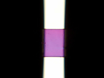
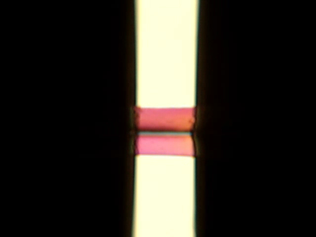
 This rheo-optics video depicts the uniaxial extensional flow deformation
of a butyl rubber at 23°C being stretched at a Hencky strain rate
of 0.3 s-1 with no back light source, just linear polarized ambient light passing through
the mid-section of the sample. Note how the entire flow field can be visualized.
This rheo-optics video depicts the uniaxial extensional flow deformation
of a butyl rubber at 23°C being stretched at a Hencky strain rate
of 0.3 s-1 with no back light source, just linear polarized ambient light passing through
the mid-section of the sample. Note how the entire flow field can be visualized.
(AVI file format: 1.55 MB)
|
 |
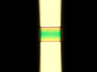
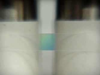
 This rheo-optics video depicts the uniaxial extensional flow deformation
of a butyl rubber at 23°C being stretched at a Hencky strain rate
of 0.3 s-1 with a linear polarized halogen white light passing through
the mid-section of the sample. Note the uniform color evolution in the sample, evidence of
the uniformity of the applied extensional deformation.
This rheo-optics video depicts the uniaxial extensional flow deformation
of a butyl rubber at 23°C being stretched at a Hencky strain rate
of 0.3 s-1 with a linear polarized halogen white light passing through
the mid-section of the sample. Note the uniform color evolution in the sample, evidence of
the uniformity of the applied extensional deformation.
(AVI file format: 0.31 MB)
|
 |
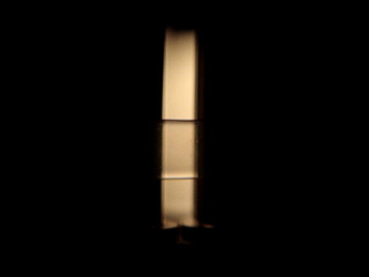
 This rheo-optics video depicts stress relaxation of a butyl rubber at 23°C after being
stretched at a Hencky strain rate of 0.3 s-1 for a total applied Hencky strain of 2.5.
Note how the stress fringes depict how the web of the sample relaxes non-uniformly from the center
of the sample towards its edges as it attempts to recoil from the applied deformation.
This rheo-optics video depicts stress relaxation of a butyl rubber at 23°C after being
stretched at a Hencky strain rate of 0.3 s-1 for a total applied Hencky strain of 2.5.
Note how the stress fringes depict how the web of the sample relaxes non-uniformly from the center
of the sample towards its edges as it attempts to recoil from the applied deformation.
(AVI file format: 2.46 MB)
|
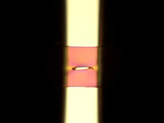 |
 This rheo-optics video depicts stress relaxation of a butyl rubber sample
with a small entrapped bubble void in its center at 23°C after being
stretched at a Hencky strain rate of 0.3 s-1 for a total applied Hencky strain of 1.
Note the stress fringe evolution around the bubble and how the bubble size remains stable during
relaxation.
This rheo-optics video depicts stress relaxation of a butyl rubber sample
with a small entrapped bubble void in its center at 23°C after being
stretched at a Hencky strain rate of 0.3 s-1 for a total applied Hencky strain of 1.
Note the stress fringe evolution around the bubble and how the bubble size remains stable during
relaxation.
(AVI file format: 2.37 MB)
|
 |
 This rheo-optics video depicts stress relaxation of a butyl rubber sample
with an identical entrapped bubble void as the sample above at 23°C after being
stretched at a Hencky strain rate of 0.3 s-1 for a total applied Hencky strain of 2.
Note how a critical strain is surpassed thereby driving bubble instability of the entrapped void and the eventual cleaving of the sample.
This rheo-optics video depicts stress relaxation of a butyl rubber sample
with an identical entrapped bubble void as the sample above at 23°C after being
stretched at a Hencky strain rate of 0.3 s-1 for a total applied Hencky strain of 2.
Note how a critical strain is surpassed thereby driving bubble instability of the entrapped void and the eventual cleaving of the sample.
(AVI file format: 1.97 MB)
|
|

|
 |
 This rheo-optics video depicts tensile elongation of a solid LLDPE sample
at 23°C stretched at a Hencky strain rate of 0.1 s-1 all the way to rupture.
This rheo-optics video depicts tensile elongation of a solid LLDPE sample
at 23°C stretched at a Hencky strain rate of 0.1 s-1 all the way to rupture.
(AVI file format: 3.44 MB)
|
|

|
With the use of a white light source, a fiber optic portal into the oven, and a high
temperature linear polarizer optical filter, rheo-optics measurements can also be made
on polymer melts within an oven chamber.
For instance, the simple experimental setup shown below depicting a model SER-HV-P01
mounted on an Anton Paar MCR host rotational rheometer
system equipped a CTD600 oven was used to capture the following
rheo-optics videos utilizing a reflector on the end
of a fiber optic cable to reflect white light off of the internal back surface of the oven
enclosure and a high-temperature linear polarizer filter mounted on the back side of the SER that was
used to polarize white light passing through the deforming sample and out the optical
window of the oven to the linear polarizer mounted on the video camera located outside
of the oven. The following videos depict parallel polarized light passing through the polymer melt
samples undergoing stretch.
|
|

|
 |
 This rheo-optics video depicts the uniaxial extensional flow deformation
of a molten HDPE sample
at a temperature of 129°C (just 0.8°C below its peak melt temperature) stretched at a
Hencky strain rate of 0.3 s-1. Note how the polymer sample remains perfectly
clear during the entire deformation, evidence that the sample is still molten. The type of
ductile fracture is also consistent with typical molten polymer behavior.
This rheo-optics video depicts the uniaxial extensional flow deformation
of a molten HDPE sample
at a temperature of 129°C (just 0.8°C below its peak melt temperature) stretched at a
Hencky strain rate of 0.3 s-1. Note how the polymer sample remains perfectly
clear during the entire deformation, evidence that the sample is still molten. The type of
ductile fracture is also consistent with typical molten polymer behavior.
(AVI file format: 0.65 MB)
|
 |
 This rheo-optics video depicts the uniaxial extensional flow deformation
of the same molten HDPE sample as above
at a temperature of 128.5°C (just 0.5°C cooler than the sample above) stretched at a
Hencky strain rate of 0.3 s-1. Note how the polymer sample becomes a bit cloudy
immediately upon deformation and gets darker as the deformation continues, evidence of a
quiescent flocculation of crystallites followed by a crystallization density increase with
increasing deformation. The state of the polymer sample is definitely solid-like as witnessed
by the type of fracture observed.
This rheo-optics video depicts the uniaxial extensional flow deformation
of the same molten HDPE sample as above
at a temperature of 128.5°C (just 0.5°C cooler than the sample above) stretched at a
Hencky strain rate of 0.3 s-1. Note how the polymer sample becomes a bit cloudy
immediately upon deformation and gets darker as the deformation continues, evidence of a
quiescent flocculation of crystallites followed by a crystallization density increase with
increasing deformation. The state of the polymer sample is definitely solid-like as witnessed
by the type of fracture observed.
(AVI file format: 0.77 MB)
|

|